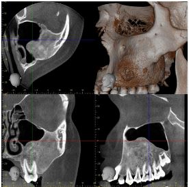
CBCT imaging of the left maxillary region shows diffuse expansion of the marrow cavity of the alveolar bone in the floor of the sinus, and diffuse sclerosis of the marrow. Appearances are compatible with fibrous dysplasia of the left maxilla.
The expansile lesion is well defined, and replaces over 50% of the maxillary antral cavity. Mild bulging of the anterior maxillary wall is noted. Superiorly, sclerotic bone abuts the inferior orbital wall, and surrounds the anterior half of the infra-orbital nerve canal. No orbital floor displacement.
Conclusion: ULQ maxillary expansile bone lesion, with well defined margins, diffuse marrow expansion showing ground glass sclerosis, and thinned but intact cortex. No aggressive or suspicious features.
The appearances are considered due to fibrous dysplasia. CBCT features do not favour an odontogenic lesion or osteoma.