For over ten years, Cavendish Imaging has been providing CBCT for craniomaxillofacial surgical applications, particularly for complex cases of skeletal abnormality and those requiring combined orthodontic/surgical management.
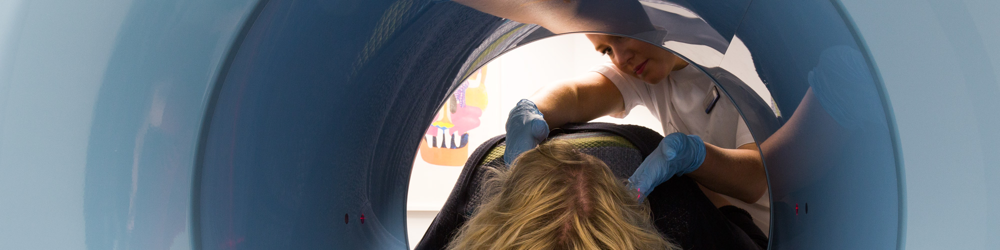
Craniomaxillofacial Surgery

The Benefits of Cavendish Cone Beam CT for CMF
Large volume CBCT scans are frequently used in planning the definitive orthognatic procedure and it is now the preferred imaging method.
A key aspect of image quality will be that we choose our CBCT scanning equipment for its image uniformity across the field of view. This means that both the virtual and 3D-printed anatomical models will be sharper than and not as noisy as what cheaper equipment other CBCT imaging centres use for their scans.
We work with both private consultants and NHS Trusts who rely on us for high imaging quality and modelling for them to achieve a very high success surgery rate in surgery and treatment and patient outcomes.
Fast, Accurate and Low Dose
Most of our CMF scans are done in upright position with a fast scanning time of approximately 10 seconds for children and 20 seconds for adults. Extra-low dose protocols will be especially used every time the emphasis for the imaging is on diagnosis only.
Expertise
Our CMF scans are reported by highly experienced UK-registered radiologists. Our in-house medical physics expertise is also at hand to help with the methodology associated with comparing pre- and post-surgery scans, with clinical and surgical accuracy measurements.
Large volume CBCT

TMJ
Individual or bilateral TMJ
An important advantage of CBCT imaging of TMJ is that it allows accurate measurements of the volume and surface of the condyle. These measurements are extremely advantageous in clinical practice when treating patients with TMJ dysfunctions (TMD). This type of scan is particularly useful in identifying and visualising osseous detail of the TMJs, including evaluation of osseous ankylosis, neoplasms, heterotopic bone growth and other abnormalities in and around the joints which might be missed in a conventional CT scan. In addition to large volume CBCT, it is worth noting that TMJ scans do not need to cover the whole facial skeleton, hence minimising further radiation to the patient. We can provide dynamic studies of the joint and in some cases register the CBCT scan against the MRI of the same region. Your patient’s scan can be done at various mandibular positions, as with conventional tomography.
Advantages of CBCT over other imaging methods for TMJ
The TMJ area is difficult to be imaged with 2D techniques due to factors like superimposition of adjacent structures and morphological variations. CBCT provides a definite advantage over these other techniques due to its low radiation dose to the patient and the ability to provide multiplanar reformation and 3D images. Indeed the complexity of the TMD demands a clear, precise and easily manipulable image of the region for effective management of the patient. When CBCT and MRI are compared, CBCT is better than MRI in detecting changes in shape (flattening, osteophyte formation or erosion) because of its superior spatial resolution and thinner slice thickness in clinical use.
Importance of diagnosing TMJ across all ages and injuries
Osteoarthritis of the TMJ is an age-related degenerative disease seen in almost 40% of patients above the age of 40 years. It causes bone changes in the TMJ like flattening, sclerosis, formation of osteophytes, erosion, resorption of the condylar head, erosion of the mandibular fossa and reduced joint space. Flattening (59%) and osteophyte (29%) are the most prevalent degenerative changes seen on CBCT. The CBCT scans play an important role in diagnosing early stages of juvenile idiopathic arthritis (JIA) in children which, when undetected, can damage facial development and cause growth alterations. Clinicians will be looking at volumetrically quantifying the TMJ damage in these patients by measuring condylar and mandibular volumes. Condylar asymmetry is very common in children with JIA. CBCT shows a wide variety of condylar destruction patterns which could be small erosions within the cortex to almost complete deformation of the head of the condyle. The TMJ is often involved in patients with multiple maxillofacial fractures. CBCT enables us to meet the patient’s needs by providing adequate information regarding the nature of fracture, its extent and relative locations of important anatomic structures.
Right-left comparison of the temporomandibular joint space

CBCT referring criteria summary
The referring criteria that we use for CBCT radiology are those of the 2012 SEDENTEX final guidelines. Incidentally, we are very proud that our own research is cited there 9 times.

It is your attention to detail and high quality scans that set you apart from your competitors
Phil Freiberger, BDS MFGDP
Quickly seen, no delay. Most welcome
E.Glover
I regularly refer patients to Cavendish Imaging for radiographs and cone beam CT. The staff are knowledgeable, helpful and also kind.
Katherine George, consultant maxillofacial and facial plastic surgeon
The operative, could not have been more helpful and nice. Also, the staff I spoke to on the phone were polite and most helpful. Very happy with the visit
R. Slotover
Thank you for being so kind and helpful throughout. I had issues getting to the appointment and your kindness and patience was much appreciated
S. Cotton
Friendly, fast, professional service. Thank you!
D. Delfavero
Walk-in clinic was very convenient and waiting time was very short.Thank you
S Griffiths
Came in with my daughter for her mouth to be scanned, both radiographers were really good with her, taking time to talk to her, explain things and put her at ease, much appreciated
E. Gowers
Overall a very prompt and efficient service
S. Foster
I was very promptly seen on arrival at the London walk-in service, the whole process required minimal time.
S. Hsu-Michel