Referring criteria: Anatomical variants prior to FESS, post FESS anatomy, mucosal inflammation, polyp grading, reactive bony sclerosis and drainage pathways.
Imaging: JMorita Accuitomo F170 and NewTom 5G
Radiation dose: Typically 150mSv (approx. 20-40% of conventional CT)
Findings: Case Studies 1-6 below illustrate the appropriateness of CBCT over CT for these referring criteria. The lower radiation dose in conjunction with the high diagnostic quality images provided by our equipment help to reinforce best clinical practice.
CBCT for Paranasal Studies

Amanda Smith BSc, Faizuddin Azizi BSc, Veronique Sauret-Jackson MPhys MSc PhD
CASE STUDY 1 – FESS planning
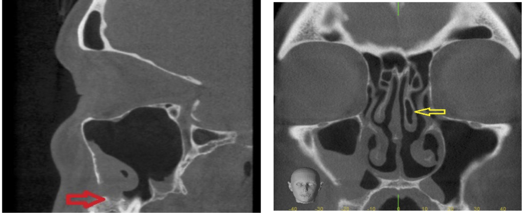
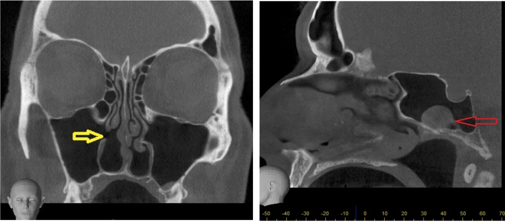
This patient presented with chronic polypoidal odontogenic sinusitis. The yellow arrow indicates a small left sided concha bullosa with no nasal septum deviation. The red arrow reveals a small 6mm x 6mm defect in the inferior aspect of the right anterolateral antral wall just above the alveolus and the right upper 5th tooth socket. The anteroinferior antral recess is lined by inflamed mucosa and there is a periosteal reaction.
CASE STUDY 2 – Sinusitis and perennial rhinitis
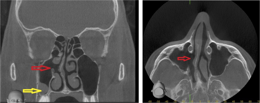
This patient had a history of septoplasty and FESS with sinus pain and headaches. The nasal septum deviates to the right slightly. There has been a right medial antrostomy (red arrows) and there is a small posteroinferior right maxillary antral mucous retention cyst (yellow arrow).
CASE STUDY 3 – Left frontal sinus fibrous dysplasia
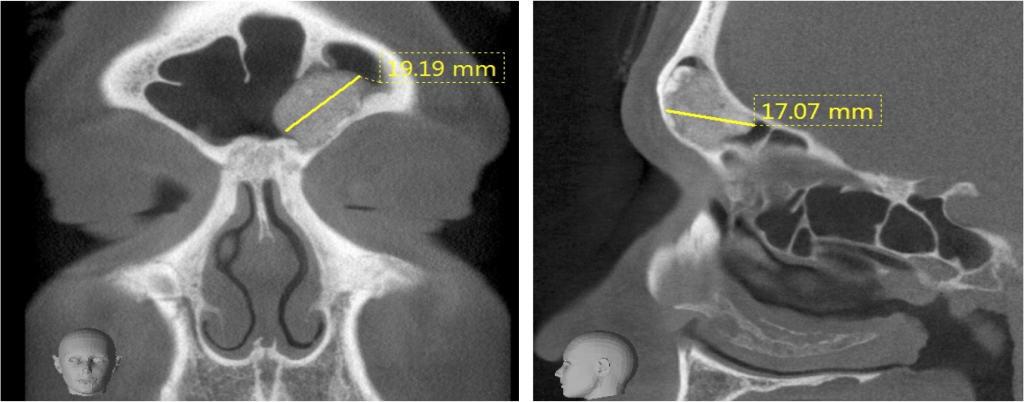
There is a well defined 17mm x 19mm lesion arising from the anterior wall and floor of the frontal sinus.
CASE STUDY 4 – Fracture nasal septum and rhinitis
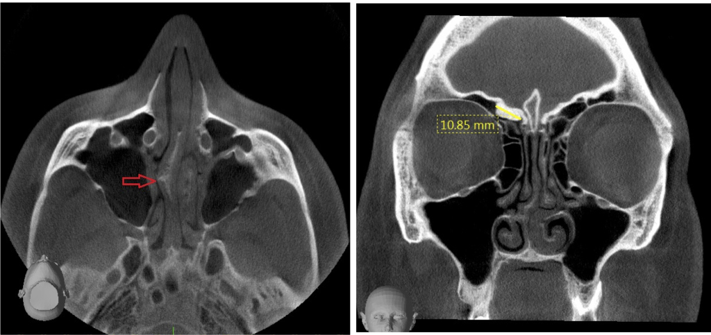
There are undisplaced, non-united fractures of the base of the nasal bone. There is a large inferior right sided septal spur shown with the red arrow. There is a 10.85mm right anterior ethmoidal osteoma.
CASE STUDY 5 – Recurrent sinus and chest infections, previous septoplasty and turbinate reduction
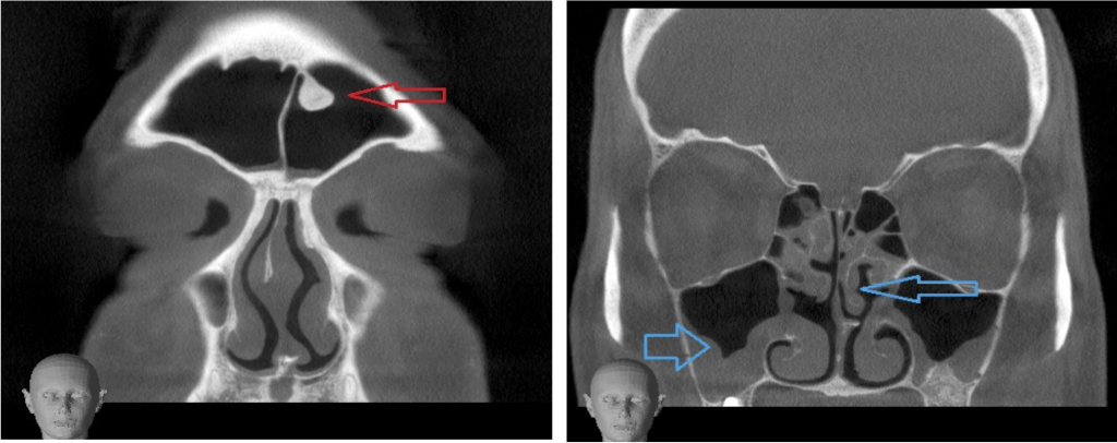
There is a defect in the medial wall of the right maxillary antrum (yellow arrow below). This is an accessory medial antral wall ostium and there is a mucous retention cyst in the left sphenoid sinus (red arrow).
CASE STUDY 6 – Sinus infection post dental treatment
There is a 4mm non-obstructive osteoma (red arrow) within the superior aspect of the left frontal sinus. There is thick lobulated sinonasal mucosal thickening of both maxillary antra and middle meati (blue arrows).
It is your attention to detail and high quality scans that set you apart from your competitors
Quickly seen, no delay. Most welcome
I regularly refer patients to Cavendish Imaging for radiographs and cone beam CT. The staff are knowledgeable, helpful and also kind.
The operative, could not have been more helpful and nice. Also, the staff I spoke to on the phone were polite and most helpful. Very happy with the visit
Thank you for being so kind and helpful throughout. I had issues getting to the appointment and your kindness and patience was much appreciated
Friendly, fast, professional service. Thank you!
Walk-in clinic was very convenient and waiting time was very short.Thank you
Came in with my daughter for her mouth to be scanned, both radiographers were really good with her, taking time to talk to her, explain things and put her at ease, much appreciated
Overall a very prompt and efficient service
I was very promptly seen on arrival at the London walk-in service, the whole process required minimal time.
