This patient injured his upper right central incisor after slipping and falling in the street.
A periapical radiograph was taken which appeared to show 2 fractures of the root. On the basis of this examination, the prognosis of the tooth seemed hopeless and the patient was sent for a 3D CBCT scan for implant planning about 2 weeks after the injury.
A scan was taken using standard settings on an Accuitomo F170 to capture a small volume of data, centred on the upper incisor teeth.
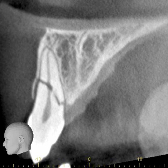
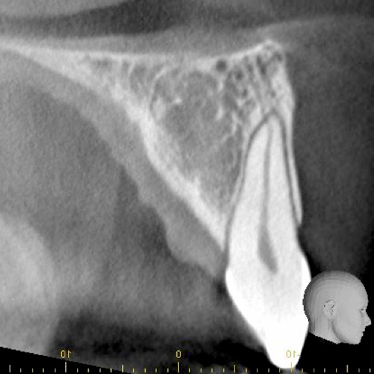
Cross sectional views through the central incisors reveal the fuller picture and permit a complete diagnosis to be made.
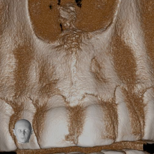
A single slightly open fracture of the root is clearly apparent. The images also reveal the vascular supply to each of the central incisor teeth (left central incisor for comparison), and a small healing fracture of the buccal plate, associated with the fractured coronal segment.
Clinically, the coronal segment was fairly firm, and symptoms from the tooth were improving.
With this new radiographic information available, the patient and referring dentist Dr Patel were able to make a fully informed decision. In this case, the plan was to keep the situation under review, deferring extraction and implant treatment for the time being.
One of our favourite anecdotes is of course that Dr Patel, specialist endodontist, has used this specific image as the front cover of the first endodontic specialist book on Cone Beam CT. It must be exceptionally good then!


